The Best anatomy of an elbow of 2022 – Reviewed and Top Rated
After hours researching and comparing all models on the market, we find out the Best anatomy of an elbow of 2022. Check our ranking below.
2,956 Reviews Scanned

- The complete joint model :The Human Body Six Major joint can meet your needs for different joint research and to provide you with a comprehensive and systematic knowledge learning and research system, is a rare auxiliary tool for your joint research.
- Medical supplies human shoulder elbow hip knee hand foot joint bone models,fine workmanship creates good item with clear shape with great detailing.so you can Lively explain the relevant knowledge to the students by the model,let students to quickly understand and master. Improve teaching quality
- Material:it is made of food grade PVC and handcrafted. fine workmanship creates good item with clear shape with great detailing.Almost true display of human joint structure. And PVC material does not have to worry about being broken by students,so It will be a great addition to your lab supplies.
- Demonstration Content: The six main joint models for the human body are suitable for medical education in the teaching of joints or ligaments or as a decoration in your laboratory more efficient to make study to the human body. So as to deepen the understanding of the structure of the human body.
- [Application] It can be used as a medical training tool, and it is also a great toy for those who want to know more about Human body structure.

- Illustrates general shoulder and elbow anatomy. Shows anterior, posterior, lateral, and superior view of the shoulder.
- Also shows the socket of shoulder joint anterior and disclocation of humerus.
- Illustrates impingement syndrome and acromioplasty.
- Shows sagittal view of the elbow, as well as supination, pronation and superior views of extension and flexion.
- Size is 20 inches by 26 inches - Flexible plastic 1.5 mil lamination - Anatomical Chart.

- Anatomy Model: GPI Anatomicals presents a scientifically accurate anatomy model of a full-size normal elbow with muscles. The model, which illustrates a right elbow from the humerus to the hand, is a great substitute for anatomy posters
- Elbow Model: The anatomy model shows all the components found in a right elbow: muscles including the biceps brachii and brachialis, bones including the humerus and ulna, ligaments including the flexor retinaculum, and nerves including the radial
- Model Specifications: The human anatomy model comes with an information card and a display base. The model measures 19" x 3" x 6", while the base measures 6-1/2" x 5". The dimensions of the information card are 8-1/4" x 6-1/4"
- Anatomy and Physiology Study Tools: This anatomy model is perfect for display in a doctor's office or a healthcare facility for effective patient education. It can also be used as a teacher's accessory for classroom demonstrations
- GPI Anatomicals: Our main focus is on prioritizing customer education. We offer different interactive models of the human body with utmost accuracy. Our models also make for one of the best gifts for medical students due to their informative nature
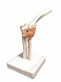
- Helps students to understand the morphology and construction of the elbow joint.
- Demonstrates all of the elbow functions and the external anatomical structures.
- Includes flexible, artificial ligaments.
- Demonstrates flexion, extension and internal/external rotation.
- Shows the humerus, radius and ulna parts of the human elbow joints.

- ▲Mini Human Joint Model Set - The 1/2 life size human joint model set of four, reduced-size joint models of the shoulder hip elbow & knee showing abduction forward, backward, inside and outside rotation, etc. Using non-toxic environmentally friendly PVC material, easy to clean and will last for years.
- ▲Model Dimensions - This model set contains four models in 1/2 life size. Hip model: 4-1/3″ x 4-1/3″ x 6-3/4″. Shoulder model: 4-1/3″ x 4-1/3″ x 4-3/4″. Knee model: 4-1/3″ x 4-1/3″ x 7″. Elbow model: 4-1/3″ x 4-1/3″ x 5-3/4″.
- ▲Medical Professional Level - The scientific human joint anatomy models were developed by medical professionals to examine various parts of the foot. Evotech Scientific provides the perfect combination of value and detail and excellent customer service to meet the needs of students and teachers.
- ▲Versatile Application - The human anatomical joint model set of shoulder knee hip elbow is suitable for doctor-patient communication. It can also be used as a teaching and study tool for medical school students, practitioners, health care professionals, schools and universities and so on.
- ▲Customer Support - If you find any problems with the human joint models, please feel free to contact our customer service and we will provide you with the most suitable solution. We are always there for you.

- Elbow Model: GPI Anatomicals presents a scientifically accurate anatomy model of a full-size normal elbow. The model, which shows the different components of a right elbow joint, is a great substitute for anatomy posters.
- Anatomy Model: The model includes the following: humerus, radius, and ulna bones; joint capsule; annular ligament of the radius, oblique cord, radial collateral, and ulnar collateral ligaments.
- Model Specifications: This human anatomy model comes with an information card and a display base. The model measures 8-1/2" x 2-1/4" x 4", while the base measures 6-1/2" x 5". The dimensions of the information card are 6-1/2" x 5-1/4".
- Anatomy and Physiology Study Tools: The anatomy model is perfect for display in a doctor's office or a healthcare facility for effective patient education. It can also be used as a teacher's accessory for classroom demonstrations.
- GPI Anatomicals: Our focus is on prioritizing customer education. We offer different interactive models of the human body with utmost accuracy. Our models also make for one of the best gifts for medical students owing to their informative nature.

- 【Elbow Joint Model】: Computer color matching, beautifully hand-painted, it is a nice model for doctors and patients to explain and compare the series of human muscle ligament strains and sprains. With the help of this model, patients can quickly understand.
- 【Teaching Tool】: Different colors are used to distinguish, and the colors are bright and easy to attract students' attention, so you can to teaching demonstration, which promotes students' understanding and increases classroom fun.
- 【PVC Material】: The anatomical model is made of odorless, durable and high quality PVC material, which is 1:1 life-size replica with a plastic base and manual for easy display, and is lightweight and easy to carry.
- 【Higher Efficient】: 1:1 life size elbow joint model is suitable for use in medical teaching when teaching skeleton, or as a decoration in your lab,making skeleton learning more efficient.
- 【Wide Application】: Applicable to schools,hospital, in physical health teaching, can be used as a teaching of physical health knowledge of the visual aids, so as to deepen the understanding of the structure of the human elbow joint.
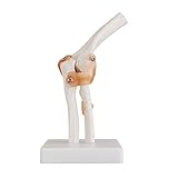
- This Functional Elbow Joint Model is made by Environmentally Friendly PVC material, very durable in use
- Environmentally friendly materials, hand-made, fine workmanship, excellent made
- This Functional Elbow Joint Model demonstrates the anatomy and mechanism of the main joints, making the patient or student understand more clearly
- Life-size, flexible rotating joint shows abduction forward tilting, backward tilting, internal and external rotation. Contains partial humerus, all ulna, and radial and joint ligament
- Comes with the base frame and can be removed

- 1:1 life size elbow joint model is suitable for use in medical teaching when teaching skeleton, or as a decoration in your lab.
- Teaching tools: It is a good model for doctors and patients to explain and compare the series of human muscle ligament strains and sprains. With the help of this model, patients can quickly understand
- Material: Made of environmentally friendly PVC, computer color matching, beautifully hand-painted. Almost true display of human elbow joint structure. Size: 20x14x15cm
- Different colors are used to distinguish , and the colors are bright and easy to attract students' attention, so you can to teaching demonstration, which promotes students' understanding and increases classroom fu
- Applicable to schools,hospital, in physical health teaching, can be used as a teaching of physical health knowledge of the visual aids, so as to deepen the understanding of the structure of the human elbow joint.

- Canine Elbow Model: GPI Anatomicals presents a scientifically accurate anatomy model of an average-sized, healthy canine left elbow joint. This model is a great substitute for animal anatomy posters
- Canine Anatomy Model: The model showcases the humerus, radius, and ulna bones, along with 6 ligaments
- Model Specifications: This canine anatomy model comes with an information card and a display base. The model measures 9-1/2" x 1-3/4" x 7-1/2", while the base measures 8-7/8" x 6-1/4". The dimensions of the information card are 6-1/4" x 8-1/4"
- Anatomy and Physiology Study Tools: The anatomy model is perfect for display in a veterinarian’s office or a healthcare facility for effective patient education. It can also be used as a teacher's accessory for classroom demonstrations
- GPI Anatomicals: Our main focus is customer education. We offer different interactive models of animal and human anatomies with the utmost accuracy. Our models also make for one of the best gifts for medical students due to their informative nature
Last update on 2025-03-18 / Affiliate links / Images from Amazon Product Advertising API
How Do You Buy The Best anatomy of an elbow?
Do you get stressed out thinking about shopping for a great anatomy of an elbow? Do doubts keep creeping into your mind? We understand, because we’ve already gone through the whole process of researching anatomy of an elbow, which is why we have assembled a comprehensive list of the greatest anatomy of an elbow available in the current market. We’ve also come up with a list of questions that you probably have yourself.
We’ve done the best we can with our thoughts and recommendations, but it’s still crucial that you do thorough research on your own for anatomy of an elbow that you consider buying. Your questions might include the following:
- Is it worth buying an anatomy of an elbow?
- What benefits are there with buying an anatomy of an elbow?
- What factors deserve consideration when shopping for an effective anatomy of an elbow?
- Why is it crucial to invest in any anatomy of an elbow, much less the best one?
- Which anatomy of an elbow are good in the current market?
- Where can you find information like this about anatomy of an elbow?
We’re convinced that you likely have far more questions than just these regarding anatomy of an elbow, and the only real way to satisfy your need for knowledge is to get information from as many reputable online sources as you possibly can.
Potential sources can include buying guides for anatomy of an elbow, rating websites, word-of-mouth testimonials, online forums, and product reviews. Thorough and mindful research is crucial to making sure you get your hands on the best-possible anatomy of an elbow. Make sure that you are only using trustworthy and credible websites and sources.
We provide an anatomy of an elbow buying guide, and the information is totally objective and authentic. We employ both AI and big data in proofreading the collected information. How did we create this buying guide? We did it using a custom-created selection of algorithms that lets us manifest a top-10 list of the best available anatomy of an elbow currently available on the market.
This technology we use to assemble our list depends on a variety of factors, including but not limited to the following:
- Brand Value: Every brand of anatomy of an elbow has a value all its own. Most brands offer some sort of unique selling proposition that’s supposed to bring something different to the table than their competitors.
- Features: What bells and whistles matter for an anatomy of an elbow?
- Specifications: How powerful they are can be measured.
- Product Value: This simply is how much bang for the buck you get from your anatomy of an elbow.
- Customer Ratings: Number ratings grade anatomy of an elbow objectively.
- Customer Reviews: Closely related to ratings, these paragraphs give you first-hand and detailed information from real-world users about their anatomy of an elbow.
- Product Quality: You don’t always get what you pay for with an anatomy of an elbow, sometimes less, and sometimes more.
- Product Reliability: How sturdy and durable an anatomy of an elbow is should be an indication of how long it will work out for you.
We always remember that maintaining anatomy of an elbow information to stay current is a top priority, which is why we are constantly updating our websites. Learn more about us using online sources.
If you think that anything we present here regarding anatomy of an elbow is irrelevant, incorrect, misleading, or erroneous, then please let us know promptly! We’re here for you all the time. Contact us here. Or You can read more about us to see our vision.
FAQ:
Q: What muscles are used in elbow?
A: Other Muscles. The pronator teres, flexor carpi radialis and extensor carpi radialis longus, three small muscles in the forearm, are also involved in elbow flexion, but not to the extent of the biceps, brachialis and brachioradialis .
Q: What muscles are around the elbow?
A: Biceps brachii: The large muscle of the upper arm flexes the arm and powerfully twists the forearm,turning the palm upward. Triceps brachii: This muscle at the back of the upper arm extends the arm and stabilizes the elbow when the hand is used for fine movements. Brachioradialis: A forearm muscle that flexes the arm at the elbow.
Q: What is inside the elbow?
A: The medial collateral ligament (MCL) of the elbow is situated on the inside of the elbow and helps to provide stability to the joint. A tear of this ligament can occur either as a sudden onset acute injury, or as a chronic, gradual onset injury through overuse.
Q: What is the anatomical name for elbow?
A: The name for the elbow in Latin is cubitus, and so the word cubital is used in some elbow-related terms, as in cubital nodes for example.